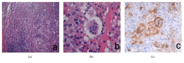
Figure 2.
Pathology of a classical case of RDD with laryngeal involvement. (a) HE staining, 60x, the patches of histiocyte proliferation form nodular zones with light staining. (b) HE staining, 300x, classical histiocytes with large nuclei and low mitotic counts; engulfed lymphocytes are present in the cytoplasm. (c) Immunohistochemical analysis revealed that the histiocytes were S-100 positive.

