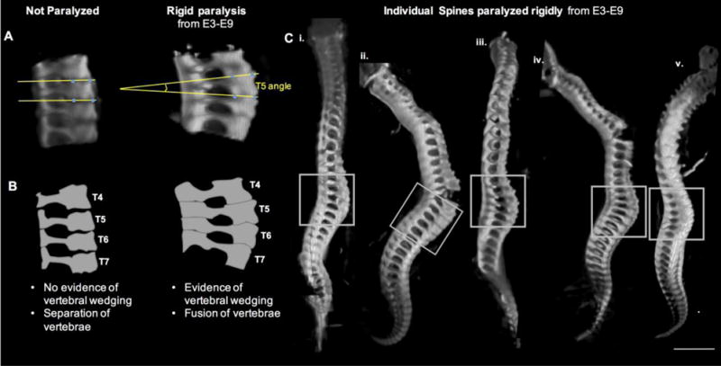
Figure 3.

Prolonged rigid paralysis led to vertebral wedging in the thoracic region. (A) Representative sagittal 3D view of thoracic spine segment (T4–T7) of control and rigidly paralyzed specimens. Yellow lines in each case show how the vertebral body angle measurements were created. (B) Schematic view of thoracic spinal segments in (A) illustrating the differences in vertebral wedging and separation of vertebrae. (C) Individual spines from 5 distinct chicks (i–v) paralyzed rigidly from E3–E9 showing evidence of vertebral wedging in the thoracic region (grey boxes). In regions of extreme curvature (indicated by arrow heads), separation at the anterior vertebral body joints occurs while the posterior spinous process joints remain intact. Scale bar 2000μm.
