Figure 1.

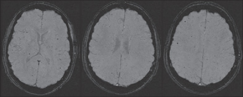
Axial susceptibility-weighted imaging images of a 16-year-old male with a history of teratology of Fallot and repair demonstrated multiple cerebral microhemorrhages.

Axial susceptibility-weighted imaging images of a 16-year-old male with a history of teratology of Fallot and repair demonstrated multiple cerebral microhemorrhages.