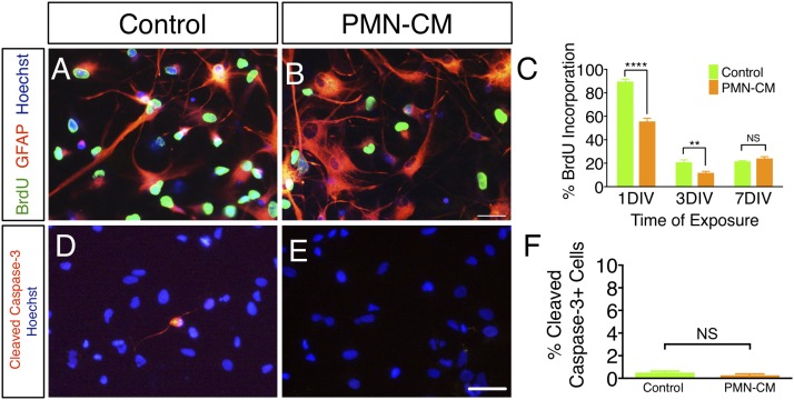
FIGURE 2.
PMN-CM decreases hNSC proliferation but does not alter hNSC death under differentiating conditions. (A and B) BrdU (green fluorescence) incorporation by hNSC treated with DM control or PMN-CM for 1 DIV. Note the increase in GFAP+ (red fluorescence) cell number and altered morphology between DM and PMN-CM. (C) PMN-CM treatment (orange bars) decreased the proportion of hNSC with BrdU incorporation versus DM control (green bars) at 1 DIV and 3 DIV, but not 7 DIV (two-way ANOVA, p < 0.0001; Bonferroni post hoc, **p < 0.01, ****p < 0.0001). (D and E) CC3 immunolabeling (red fluorescence) in hNSC exposed to DM control or PMN-CM for 1 DIV. Hoechst (blue fluorescence) indicates total nuclei. (F) PMN-CM treatment (orange bar) did not alter CC3+ cells proportion versus DM control (green bars). n = 2 technical replicates and n = 3–4 biological replicates/condition. Mean ± SEM. Scale bar, 50 μm. Student t test, p > 0.05 (NS).

