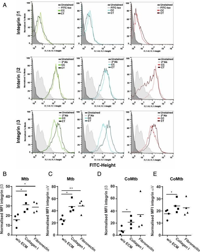
FIGURE 3.
Surface expression of β3 and αV integrin subunits in M. tuberculosis–stimulated monocytes adherent to ECM proteins. Monocytes cultured in the presence or absence of type I collagen or fibronectin were stimulated for 24 h with CoMtb (1:5 dilution), and CoMCont as control, or directly infected with M. tuberculosis (MOI = 1). Cells were fixed, blocked, and stained with FITC-conjugated anti-integrin β1or primary anti-integrin β2, β3, or αV Abs and secondary FITC-conjugated anti-mouse IgG Ab. Secondary Ab alone or FITC-conjugated IgG1 isotype Ab were used as controls. (A) Histograms of integrin subunits β1, β2, and β3 for control and CoMtb-stimulated monocytes show an increase in β3 expression with CoMtb stimulation. MFIs of integrin subunits (B) β3 and (C) αV in monocytes stimulated with M. tuberculosis, and MFIs of (D) β3 and (E) αV in monocytes stimulated with CoMtb, in the presence or absence of matrix components (n = 4). MFIs were normalized to baseline MFIs of respective controls (control media or CoMCont). (B)–(E) show data points and means. *p < 0.05.

