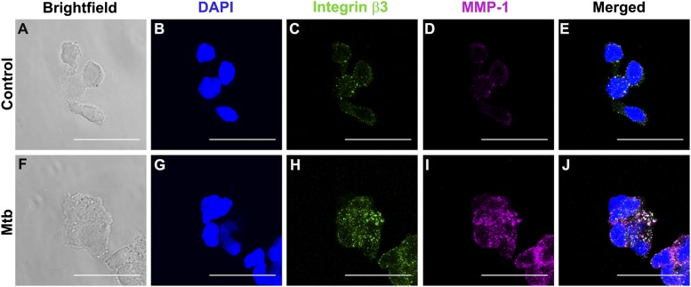
FIGURE 6.
Integrin β3 colocalizes with MMP-1 in M. tuberculosis infection. Monocytes seeded in collagen-coated chamber slides were infected with M. tuberculosis (MOI = 1). Cells were fixed, blocked, and stained with DAPI for nucleic acids (blue), integrin αVβ3 (green), and MMP-1 (magenta). (A) and (F) show brightfield and (B)–(D) show fluorescence of control monocytes and (G–I) fluorescence of M. tuberculosis–infected monocytes. (E) and (J) are merged fluorescence images. White color corresponds to areas of integrin and MMP colocalization. Scale bar, 25 μm.

