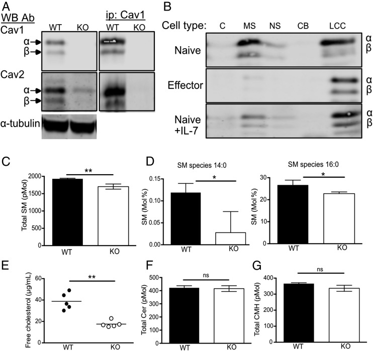
FIGURE 1.
Cav1 is localized within soluble and insoluble membrane fractions and regulates cholesterol and lipid composition. (A) Cav1 and Cav2 protein expression was compared between WT and Cav1-KO CD8 T cell lysates by Western blot with anti-Cav1 and Cav2 Ab. Cell lysates were immunoprecipitated with anti-Cav1 and Western blots were probed sequentially with anti-Cav1 and Cav2 Ab. (B) Cell lysates were separated into subcellular fractions (C, cytoplasmic; MS, membrane soluble; NS, nuclear soluble; CB, chromatin bound; LCC, lipid-cytoskeletal complexes) and analyzed by Western blot with anti-Cav1 Ab. (C) Total sphingomyelin content in WT versus Cav1-KO CD8 T cells. (D) Sphingomyelin species 14:0 and 16:0 are represented as a percentage of total sphingomyelin. (E) Free cholesterol from WT and Cav1-KO CD8 T cells is representative of one of three independent experiments from a total of 12 biological samples of each genotype. (F) Total ceramide and (G) ceramide monohexamide content. Lipidomics data are pooled from three biological repeats. Data are shown as mean + SD. *p ≤ 0.05, **p ≤ 0.01 (Student t test). ns, not significant.

