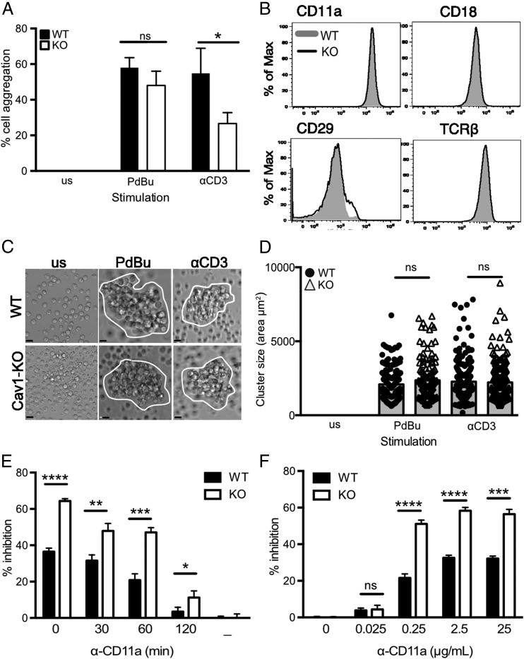
FIGURE 6.
Impaired LFA-1–mediated CD8 T cell homotypic aggregation in the absence of Cav1. (A) Purified Cav1-WT (filled bars) and Cav1-KO (open bars) OT-1 CD8 T cells were activated with 50 ng/ml PdBu or 1 μg/ml anti-CD3 for 60 min and assessed for cell–cell aggregation. Data are shown as a mean ± SEM of data representing one of five independent experiments with each count performed in triplicate on two to four mice per group. (B) Expression levels of the indicated surface proteins in WT (gray shaded) and Cav1-KO (black line) naive OT-1 CD8 T cells. Data are representative of 25 mice per group. Naive CD8 T cells were cultured for 24 h and then (C) imaged and (D) assessed for cluster size using Volocity software. Data are pooled from two independent experiments with a minimum of 100 cells per condition (mean and SD). Scale bars, 5 μm. (E) Anti–LFA-1 blocking mAb was added at various time points or (F) at various concentrations, following 1 μM N4 peptide stimulation of Cav1-WT and Cav1-KO CD8 T cells. Inhibition of CD69 upregulation (percentage inhibition) was measured at 3 h. Data are representative of one of three independent experiments. *p ≤ 0.05, **p ≤ 0.01, ***p ≤ 0.001, ****p = 0.0001 (Student t test); ns, not significant.

