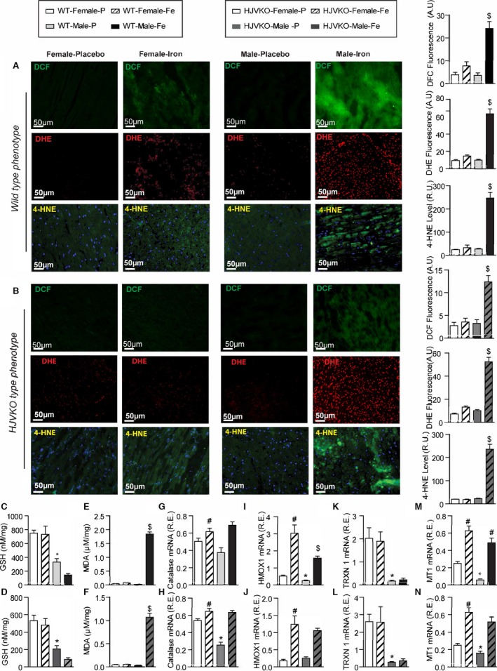
Figure 3.

Female mice are protected against iron‐overload‐induced oxidative stress. Representative dichlorodihydrofluorescein (DCF) (green), dihydroethidium (DHE) fluorescence (red), 4‐hydroxynonenal (4‐HNE) immunofluorescence (green), and quantification showing a relative lack of iron‐induced myocardial oxidative stress in female WT (A) and HJVKO (B) mice, whereas iron overload resulted in increased oxidative stress in male mice. Biochemical analysis of myocardial reduced glutathione (GSH) (C and D) and lipid peroxidation product, malondialdehyde (MDA) (E and F) levels in WT (C and E) and HJVKO hearts (D and F) showing increased oxidative stress in male mice in contrast to unchanged oxidative stress in female mice in response to iron overload. Myocardial gene expression analysis using Taqman real‐time PCR showing sex‐specific and iron‐overloaded related alteration in mRNA expression for catalase (G and H), heme oxygenase 1 (HMOX1) (I and J), thioredoxin 1 (TRXN1) (K and L), and metallothionein 1 (MT1) (M and N) in WT and HJVKO mice, respectively; n=4 for histology; n=8 for gene expression and biochemical analyses. HJVKO indicates hemojuvelin‐null; WT, wild‐type. *P<0.05 for effect of sex; # P<0.05 for effect of iron; $ P<0.05 for interaction.
