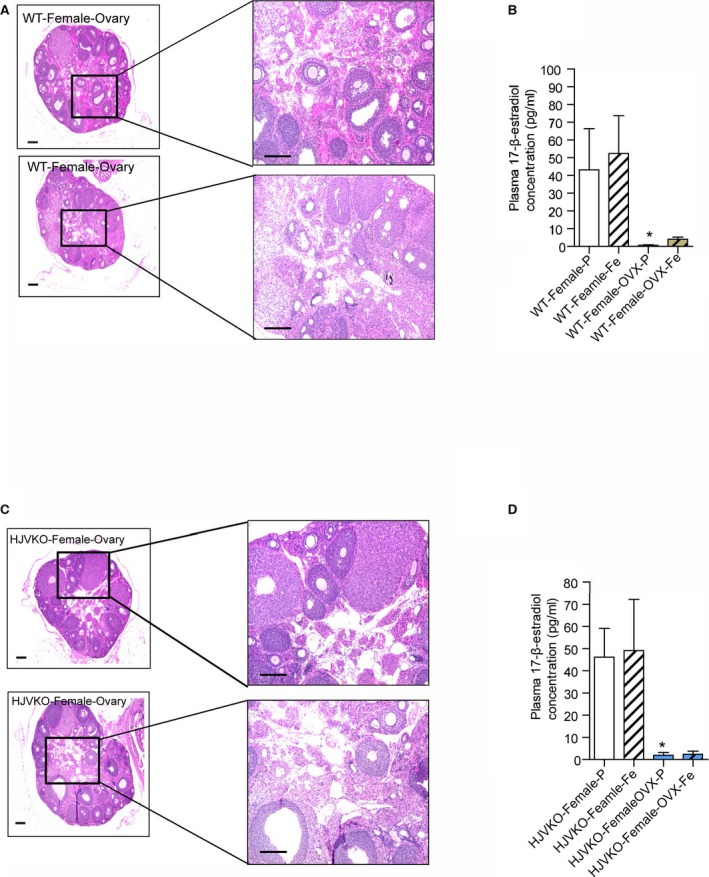
Figure 8.

Histological analysis of ovaries and plasma 17β‐estradiol levels in WT mice and HJVKO mice. Hematoxylin and eosin staining of surgically removed ovaries from a WT (A) and HJVKO (C) mouse and plasma 17β‐estradiol levels in WT (B) and HJVKO (D) mice illustrating the effectiveness of ovariectomy (OVX). Scale bars represent 250 μm (left) and 50 μm (right); n=8 per group. HJVKO indicates hemojuvelin‐null; WT, wild‐type. *P<0.05 for effect of OVX.
