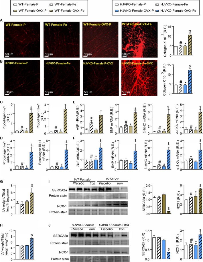
Figure 14.

Exacerbation of pathological myocardial remodeling in iron‐overloaded ovariectomized (OVX) female mice. Picro‐sirius red (PSR) staining and quantification of myocardial fibrosis (A and B) and gene expression of procollagen type Iα1 and procollagen type IIIα1 (C and D) in wild‐type (WT) and hemojuvelin‐null (HJVKO) females clearly demonstrating that OVX potentiates iron‐overload‐mediated myocardial fibrosis. Expression of disease markers, atrial natriuretic factor (ANF), brain natriuretic peptide (BNP), β‐myosin heavy chain (β‐MHC), and α‐skeletal actin (α‐SkA) in WT (E) and HJVKO (F) mice illustrating pathological myocardial remodeling in iron‐overloaded OVX female hearts. Morphometric assessment of hypertrophy showing increased LV weights in iron‐overloaded OVX female WT (G) and HJVKO (H) hearts. Western blot analysis and quantification clearly showed significant downregulation in myocardial sarco/endoplasmic reticulum Ca2+ ATPase 2a (SERCA2a) and sodium‐calcium exchanger 1 (NCX1) levels in female WT (I) and HJVKO (J) iron‐overloaded hearts following OVX. R.E. indicates relative expression; R.F., relative fraction; R.R., relative ratio; n=8 for gene expression analysis; n=4 for histology and Western blot analyses. *P<0.05 for effect of OVX; # P<0.05 for effect of iron; $ P<0.05 for interaction.
