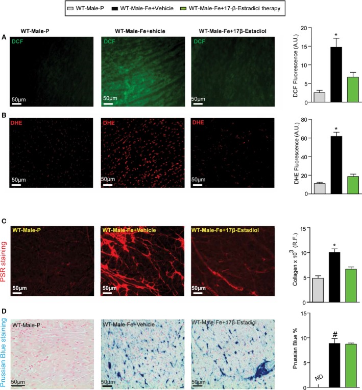
Figure 16.

Reduction in myocardial oxidative stress and fibrosis in male iron‐overloaded mice in response to 17β‐estradiol therapy. Representative dichlorodihydrofluorescein (DCF) fluorescence (green) (A) and dihydroethidium (DHE) fluorescence (red) (B) and quantification showing a marked suppression of myocardial oxidative stress in male WT iron‐overloaded mice in response to 17β‐estradiol therapy, with a similar change seen in myocardial fibrosis as determined by picro‐sirius red (PSR) staining (C) without suppression of myocardial iron deposition as illustrated by Prussian blue staining (D); n=4 for histology analysis. R.F. indicates relative fraction; WT, wild‐type. *P<0.05 compared with all the groups; # P<0.05 compared with the placebo group.
