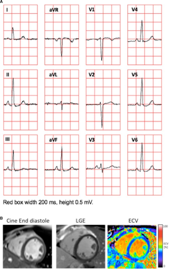
Figure 4.

Example of a 24‐year‐old woman illustrating the coexistence of normal QRS amplitudes on the ECG in patients with high left ventricular mass index (LVMI) and increased myocardial extracellular volume fraction (ECV). A, The 12‐lead ECG. The Sokolow‐Lyon index was 2.8 mV, Cornell voltage 0.3 mV, and the 12‐lead voltage was 14.7 mV. B, An end‐diastolic cine image from a midventricular short‐axis slice, which was part of the short‐axis stack from which the LVMI (60 g/m2) was measured (left), a corresponding late gadolinium enhancement (LGE) showing absence of focal abnormalities (middle), and a corresponding ECV image showing diffusely increased ECV (30%) consistent with diffuse myocardial fibrosis (right).
