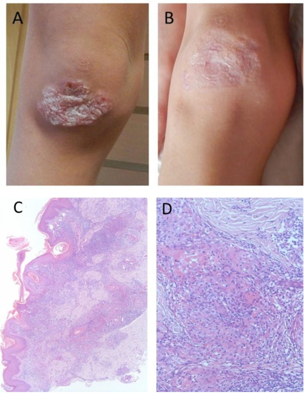
Figure 1.

Macroscopic and microscopic images of granulomatous lesion in a boy with Nijmegen Breakage Syndrome: (A) at referral, after multiple courses of antibiotics and topical steroids, (B) after conditioning for hematopoietic stem cell transplantation, (C) skin histology demonstrating perivascular and diffuse, mixed-cell infiltrate in superficial and deep dermis. H&E stain, 40× magnification, (D) skin histology demonstrating palisaded inflammatory infiltrates consisting of histocytes, lymphocytes, and neutrophils, associated with basophilic alterations of collagen. H&E stain, 400× magnification.
