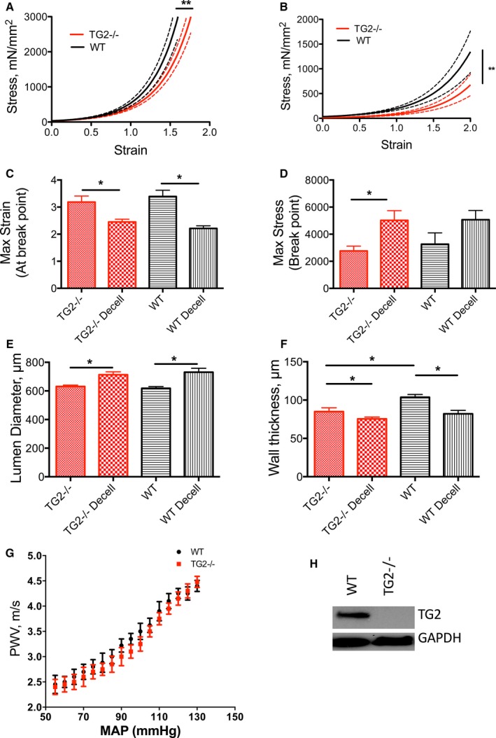
Figure 4.

TG2 contributes to aortic modulus. A, Decellularized and (B) intact aortic segments from WT mice were significantly stiffer than those from TG2−/− mice; solid lines represent arithmetic mean and dotted lines represent SE of measurement (*P<0.05). C, Maximum strain at sample rupture was lower in the decellularized segments than in intact segments and similar between genotypes (*P<0.05). D, Maximum stress was higher in the decellularized segments than in intact aorta for the TG2−/− genotype but not the WT genotype. E, Lumen diameters were larger in decellularized segments than in intact segments for WT aorta (*P<0.05). F, Walls were significantly thinner in decellularized samples than in intact samples, in WT aorta (n=12 per group; *P<0.05). G, Pulse wave velocity (PWV)–mean arterial pressure (MAP) correlation was similar in 12‐ to 14‐week‐old WT and TG2−/− mice (n=8). H, Representative Western blot showing that TG2 protein was expressed in aorta from WT but not TG2−/− mice; GAPDH was used as loading control. TG2 indicates tissue transglutaminase; WT, wild type.
