Figure 4.

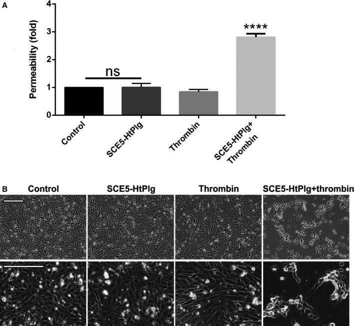
Brain microvascular endothelial cells were cultured to confluence in permeable Transwell inserts and incubated for 6 hours with anti–glycoprotein IIb/IIIa single‐chain antibody–human thrombin‐activatable microplasminogen (SCE5‐HtPlg; 100 nmol/L) only, thrombin only (2.5 U/mL), and SCE5‐HtPlg (100 nmol/L) with thrombin (2.5 U/mL). A, Permeability was measured by fluorescein isothiocyanate‐BSA passage through the monolayers over 1 hour and presented as mean±SEM values of permeability normalized to untreated controls (n=3, ****P<0.0001, nonsignificant [ns]). B, Representative phase‐contrast images of brain endothelial cells 12 hours after various treatments. Prominent gaps and morphology changes are observed in the combined treatment group, but not in cells treated with (nonactivated) SCE5‐HtPlg alone.
