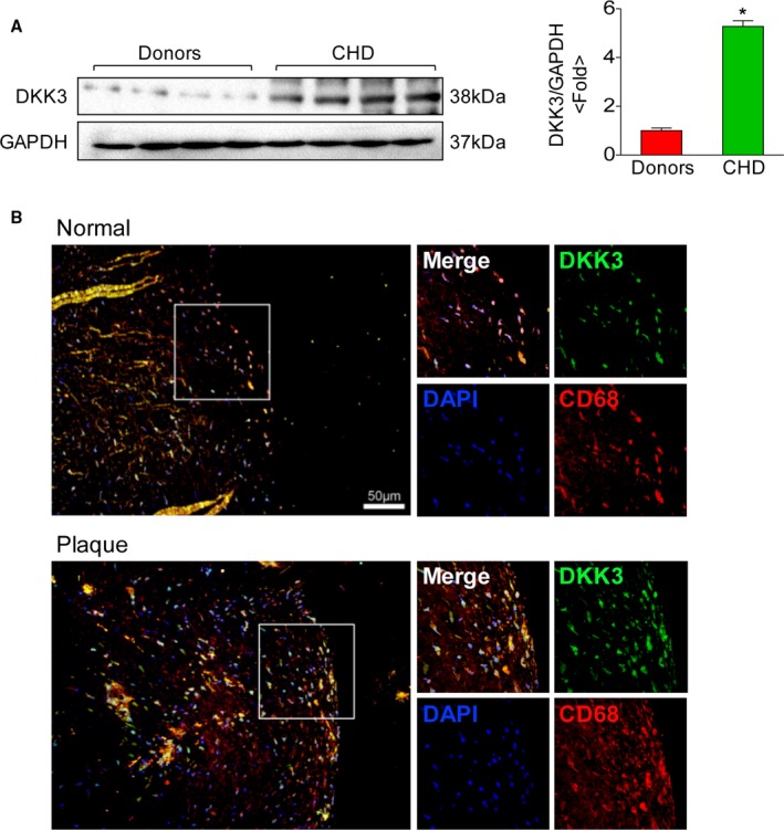
Figure 7.

Expression of DKK3 in human atheromatous lesions. A, Western blotting analysis of DKK3 protein levels in the right coronary artery in humans. The expression levels were normalized to GAPDH and quantified. n=4. *P<0.05. B, Representative images showing double‐immunofluorescence staining of human coronary arteries for DKK3 (green) and macrophages (red). Scale bar=50 μm. DKK3 indicates Dickkopf‐3.
