
Figure 2.
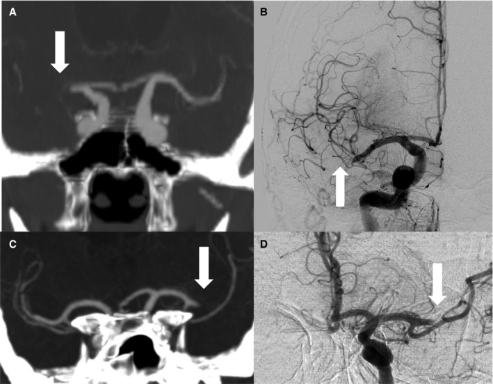
Thrombus migration vs stable clot position on sequential CTA/DSA. Upper row (A+B), case with direct evidence of thrombus migration; (A) coronary CTA reformation with 10 mm maximum intensity projection shows proximal MCA occlusion on the right side (arrow); (B) initial DSA runs before recanalization revealed significant thrombus dislocation (see arrow) to the distal M1‐segment/Truncus inferior (migration group A); lower row (C+D), case with stable clot position; (C) coronary CTA reformation with 10 mm maximum intensity projection displays proximal thrombus end after MCA bifurcation (arrow); (D) unchanged thrombus position on first DSA runs before recanalization (arrow). CTA, computed tomographic angiography; DSA, digital subtraction angiography; MCA, middle cerebral artery.