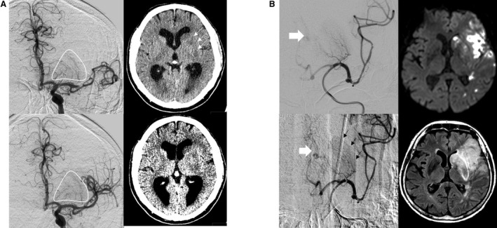
Figure 5.

Signs of preinterventional infarction. A, Distal MCA occlusion with clear evidence of preinterventional infarction of the striatocapsular region. First DSA runs prior to endovascular treatment revealed full perfusion of all perforators (compare pre‐ and postinterventional DSA, left side upper and lower row). Preinterventional CT before DSA revealed demarcation of striatocapsular infarcts (see arrows) and hypodensities on “stroke window” settings (right side lower row). B, Case with distal MCA occlusion and clear evidence of preinterventional infarction of the striatocapsular region. First DSA runs prior to endovascular treatment revealed full perfusion of all MCA perforators but basal ganglionic blush (see black arrows, left side lower row) and early venous drainage (left side, white arrows), suggesting an already occurred infarction. Postinterventional DWI (right side upper row) and FLAIR‐sequence revealed complete striatocapsular infarct corresponding to a complete occlusion of all MCA perforators. CT, computed tomography; DSA, digital subtraction angiography; DWI, diffusion weighted imaging; FLAIR, fluid attenuation inversion recovery; MCA, middle cerebral artery.
