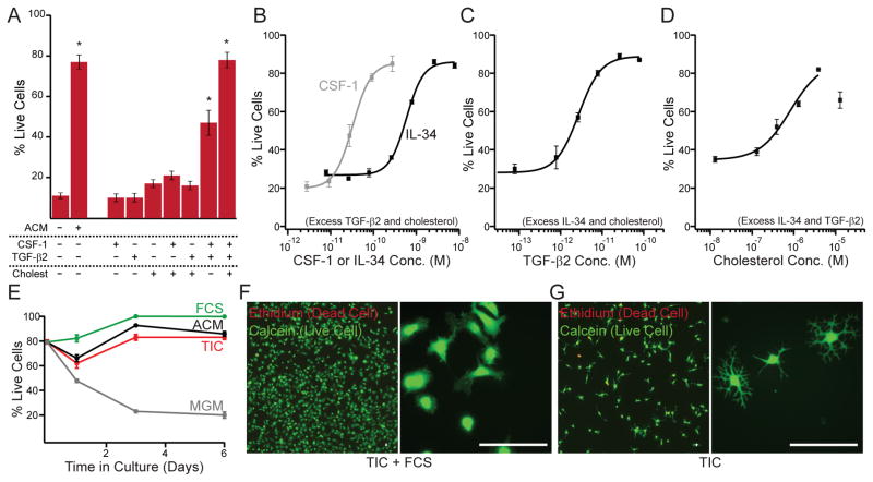
Figure 2. The combination of TGF-β, CSF-1/IL-34, and cholesterol supports survival of ramified microglia in defined medium.
(A) The combination of CSF-1 (10 ng/mL), TGF-β2 (2 ng/mL), and cholesterol (1.5 μg/mL) facilitates survival of primary rat microglia as well as ACM (20 μg/mL). Individual components or pairwise combinations were not sufficient for robust survival (5 div). Some bars are replotted from Fig. 1C for clarity. (B) Dose-response analysis of CSF-1 and IL-34 when supplemented with TGF-β2 (2 ng/mL) and cholesterol (1.5 μg/mL). (C) Dose-response analysis of TGF-β2 when supplemented with IL-34 (100 ng/mL) and cholesterol (1.5 μg/mL). (D) Dose-response analysis of cholesterol when supplemented with IL-34 (100 ng/mL) and TGF-β2 (2 ng/mL). Lines in (B–D) describe Hill equation fits. (E) Time course of microglial survival in base medium (MGM, gray), ACM (black), TIC (red) or TIC+10%FCS (green). (F, G) Calcein/ethidium-labeled microglia grown in TIC supplemented with 10% FCS (F) or TIC alone (G) illustrate serum-induced proliferation (low magnification, left) and altered morphology (high magnification, right). Averages are mean ± sem. Scale bars = 40 μm. * P < 0.001 one-way ANOVA across all groups with Dunnett’s comparison to no additive condition.

