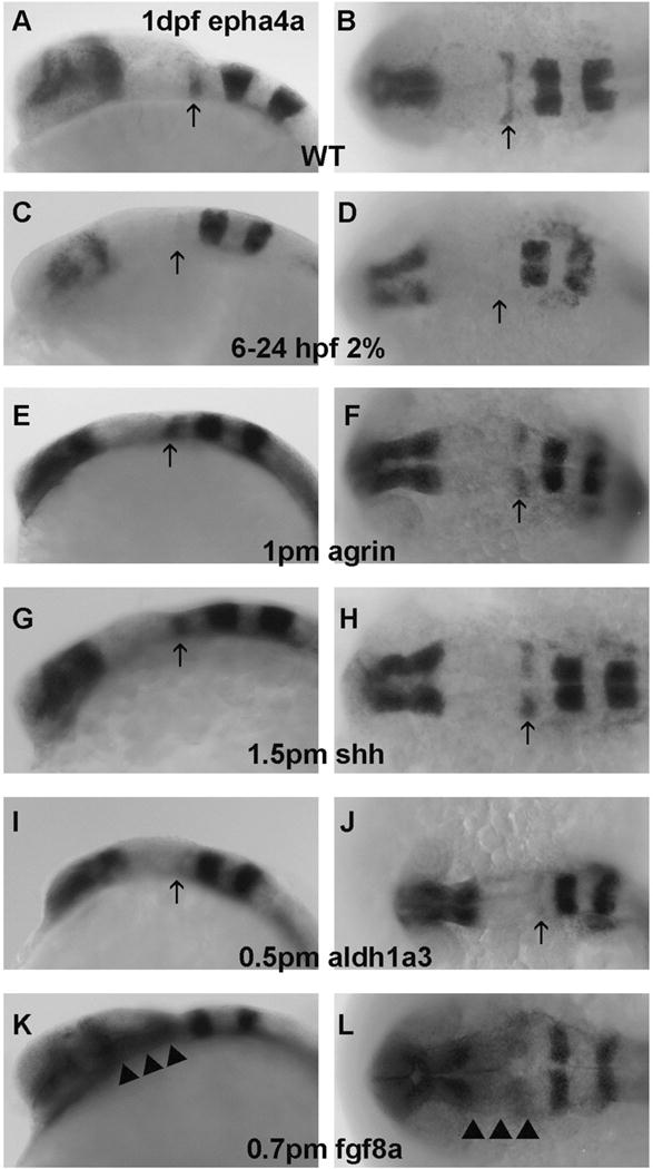
Figure 3. EphA4a mRNA expression in R1 boundary is selectively decreased in embryos exposed to chronic ethanol treatment or aldh1a3 MO.

A–B, WT; C–D, embryos exposed to 2% ethanol from 6–24 hpf; E–F, embryos injected with 1 pmol agrin MO; G–H, embryos injected with 1.5 pmol shh MO; I–J, embryos injected with 0.5 pmol aldh1a3 MO; K–L, embryos injected with 0.7 pmol fgf8a MO. A,C,E,G,I, and K embryos are lateral views; B,D,F,H,J, and L embryos are dorsal views. Arrows indicate R1 boundary expression. Note that ephA4a expression in R1 decreased with ethanol exposure (C–D), aldh1a3 MO(I–J) or fgf8a MO (K,L). Ectopic epha4a expression in posterior midbrain (indicated by arrowheads) was only found in fgf8a morphants (K–L).
