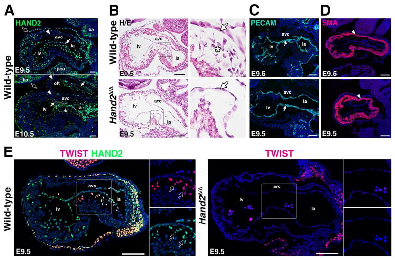
Figure 2. AVC cardiac cushion agenesis in Hand2-deficient mouse embryos.
(A) Distribution of the endogenous HAND23xF protein (using anti-FLAG antibodies, green fluorescence) during in mouse embryonic hearts at E9.5 and E10.5. Representative sagittal sections are shown. White arrows: endocardium; white arrowheads: myocardium; white asterisks: HAND2 expressing AVC cardiac cushion mesenchymal cells; black arrows: epicardium. Scale bars: 50μm. (B) Hematoxylin/Eosin staining reveals the absence of delaminating endocardial cells with mesenchymal characteristics (white asterisks) in the AVC of Hand2-deficient mouse embryos. White arrow: endocardium. Scale bars: 100μm. (C, D) Detection of the platelet endothelial cell adhesion molecule (PECAM) in the endocardium and smooth muscle actin (SMA) in the AVC myocardium of wild-type and Hand2-deficient embryos. White arrow: endocardium, white arrowhead: myocardium (panels A to D). (E) Colocalization of the HAND23xF (green fluorescence) with TWIST1 transcriptional regulators (red fluorescence) in the AVC of wild-type (Hand23xF/3xF) and Hand2-deficient (Hand2Δ/Δ) embryos at E9.5. Colocalization is detected in delaminating mesenchymal cells (indicated by arrows, left panel), which are missing from the mutant AVC (right panel). Scale bars: 100μm. avc: atrioventricular canal, ba: branchial arches, la: left atrium, lv: left ventricle, oft: outflow tract, peo: proepicardial organ.

