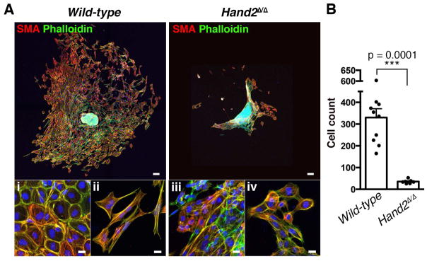
Figure 3. Hand2-deficient AVC endocardial cells fail to initiate EMT and mesenchymal cell migration.
(A) Smooth muscle actin (red) and F-actin (green) distribution in cells that have migrated into the matrix from AVC explants of wild-type and Hand2-deficient embryos after 72hrs in culture. Scale bars: 100μm. Bottom panels show cells in proximity to the explant (i and iii) and at the far edge of migration (ii and iv). Scale bars: 20μm. (B) Quantification of the numbers of cells that migrated into the matrix from wild-type (n=10) and Hand2-deficient AVC explants (n=7). The mean ± SD (p=0.0001, Mann-Whitney test) and all individual data points are shown.

