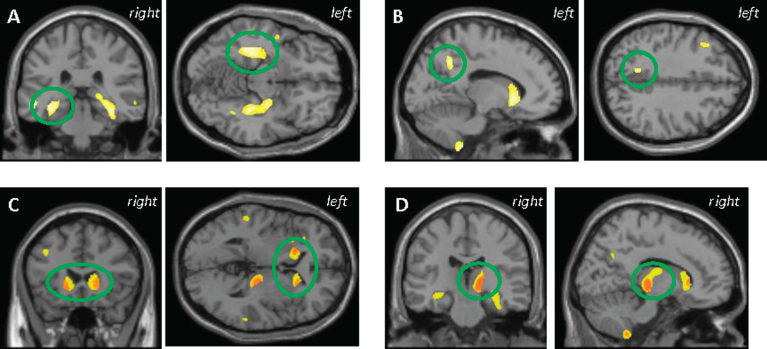
Fig.1.
VBM analysis in all participants showed higher plasma tau was correlated with lower grey matter density (GMD) in the (A) medial temporal lobe, (B) precuneus, (C) striatum, and (D) thalamus. C and D represent the anatomic overlap (orange) of regions of GM atrophy associated with higher plasma tau using only age, sex, APOE ɛ4 status, and total intracranial volume as covariates (yellow) and with the addition of diagnosis as a covariate (red). Results are displayed at p < 0.001 (uncorrected) and at a threshold (k) of 100 voxels.

