Figure 5.

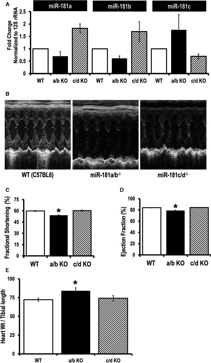
Examination of cardiac function in miR‐181a/b−/− and miR‐181c/d−/− mice. A, Quantitative polymerase chain reaction (SYBR kit) analysis of miR‐181a, b, and c expression in total RNA from the heart tissues of miR‐181a/b−/− (a/b KO), and miR‐181c/d−/− (c/d KO) mice. The miRNA expression was normalized to mitochondrial 16S rRNA. B, Two‐dimensional M‐mode and Doppler echocardiography were performed on nonanesthetized 12 week old mice: wild type (WT C57BL6) mice (left), miR‐181a/b−/− mice (middle panel), and miR‐181c/d−/− mice (right). C, Percentage fractional shortening and (D) ejection fraction were calculated using the software of the echocardiography instrument (n=8). E, Signs of hypertrophy (heart weight/tibial length) were measured (n=5). *P<0.05 vs WT.
