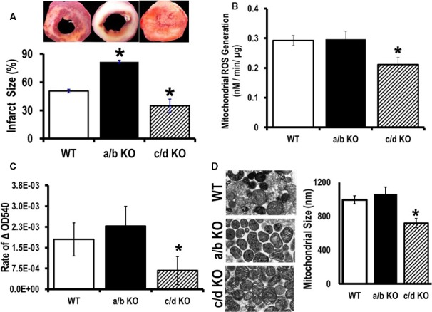
Figure 8.

Cardiopretective effect of microRNA (miR)‐181c/d−/− from ischemia/reperfusion injury. A, Infarct size was calculated after 20 minutes of global ischemia, followed by 2 hours of reperfusion from 3 groups of animals: wild type (WT), miR‐181a/b−/−, and miR‐181c/d−/−. B, Rate of reactive oxygen species (ROS) generation and (C) mitochondrial swelling was measured from isolated heart mitochondria from the 3 groups of mice: WT, miR‐181a/b−/− (a/b KO), and miR‐181c/d−/− (c/d KO), using glutamate/malate as a substrate. D, Electron microscopy of mitochondria isolated from mouse hearts. Representative pictures of the 3 groups of animals, WT, miR‐181a/b−/− (a/b KO), and miR‐181c/d−/− (c/d KO), is shown on the left side. Transmission electron microscope measurement of average longitudinal mitochondrion size is represented by a bar graph on the right side. *P<0.05 vs WT (n=6).
