
Figure 2.
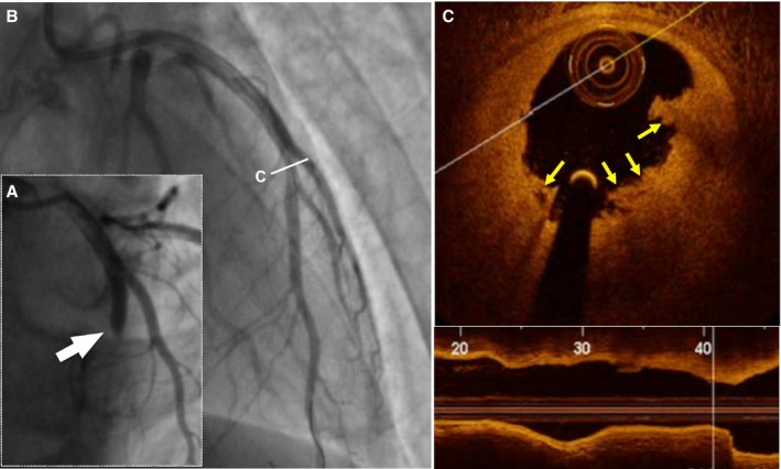
Representative optical coherence tomography (OCT) images of plaque erosion. A 28‐year‐old man with ST‐segment elevation myocardial infarction was admitted after 2 hours of chest pain. Results from index angiogram showed total occlusion in the mid–left anterior descending artery (LAD; white arrow, A). Angiogram results after thrombus aspiration showed a mild stenosis in the mid‐LAD (B). Cross‐sectional image of the culprit lesion shows residual white thrombus without evidence of ruptured fibrous cap (yellow arrows, C). The patient was treated with medical therapy without stent implantation.