
Figure 3.
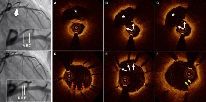
Representative optical coherence tomography (OCT) images of plaque rupture. A 60‐year‐old woman with ST‐segment elevation myocardial infarction was admitted after 11 hours of chest pain. Results from diagnostic angiogram showed total occlusion in the proximal left anterior descending artery (LAD). Angiogram results after thrombectomy demonstrates dissection lesion in the proximal LAD (white arrow, left upper panel). Cross‐sectional OCT images of the culprit lesion show disrupted fibrous cap (white arrows) and cavity (asterisk) formation (A through C). The patient was treated with a drug‐eluting stent (3.0×18 mm). Poststenting OCT revealed in‐stent thrombus (red arrows, D), malapposed struts (white arrows, E), and stent protrusion (yellow arrows, F).