
Figure 6.
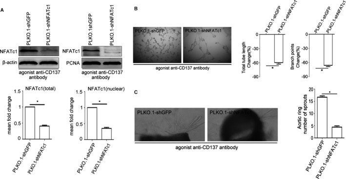
NFATc1 gene suppressing inhibited angiogenesis induced by CD137 signaling in vitro. A, NFATc1 knockdown stable cell lines (PLKO.1‐shNFATc1, PLKO.1‐shGFP as control) were treated with agonist anti‐CD137 antibody. Western blot reveals the protein level of NFATc1 in stable cell lines. Average densitometric values normalized against those of internal control from 3 independent experiments are shown in the bar graph. NFATc1 (total) median=0.40, IQR=0.09, NFATc1 (nuclear) median=0.36, IQR=0.11, n=3. B and C, Suppression of NFATc1 reduced agonist anti‐CD137 antibody‐mediated HUVEC Matrigel tube formation and decreased agonist anti‐CD137 antibody‐induced vessel outgrowth in aortic ring assays. Tube length: PLKO.1‐shNFATc1 median=−58.23, IQR=5.94, n=5. Branch points: PLKO.1‐shNFATc1 median=−66.83, IQR=5.77, n=5. Aortic ring PLKO.1‐shGFP median=16.5, IQR=1.93, PLKO.1‐shNFATc1 median=4.33, IQR=1.18, n=5. All images shown are representative and values are expressed as mean±SEM *P<0.05. HUVEC indicates human umbilical vein endothelial cell; IQR, interquartile range; NFATc1, nuclear factor of activated T cells 1; PCNA, proliferating cell nuclear antigen.; PLKO.1‐shGFP, PLKO.1‐short hairpin GFP; PLKO.1‐shNFATc1, PLKO.1‐short hairpin NFATc1