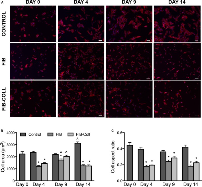
Figure 2.

VICs cultured in FIB or FIB‐Coll conditions exhibit a more elongated and spindled morphology compared with control VICs. A, Staining for phalloidin (red), nuclei are counterstained blue. Scale bar represents 100 μm. Quantification of (B) cell shape and (C) aspect ratio for VICs cultured in control, FIB, or FIB‐Coll conditions. *P<0.01 compared with control VICs at the same time point. ^ P<0.01 compared with day 4 VICs cultured in the same condition. n=6. FIB indicates fibroblast media formulation; FIB‐Coll, fibroblast media formulation with culture on collagen coatings; VIC, valvular interstitial cell.
