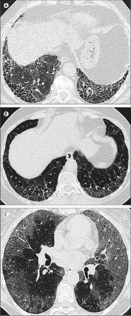
Figure 1. High-resolution CT imaging of interstitial pneumonia patterns: different diagnostic categories.
(A) Usual interstitial pneumonia pattern: axial chest high-resolution CT image taken at the level of the lower lobes, depicting multilayered subpleural honeycombing without evidence of features inconsistent with a usual interstitial pneumonia pattern. (B) Possible usual interstitial pneumonia: axial chest high-resolution CT image taken at the level of the lower lobes, depicting bilateral symmetrical reticular abnormalities containing areas of traction bronchiectasis, but no clear evidence of subpleural honeycombing. Although there are admixed areas of ground glass opacification, the predominant abnormality is of reticulation. (C) Inconsistent with usual interstitial pneumonia: axial chest high-resolution CT image taken below the level of the carina, depicting diffuse ground glass abnormalities in a predominantly peripheral distribution. Mild traction bronchiectasis is shown in the left upper lobe and right upper lobe, indicating that a proportion of the ground glass abnormality represents fibrosis.

