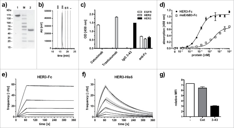
Figure 1.

Biochemical characterization and antigen binding of IgG 3–43. (a) SDS-PAGE analysis (Coomassie stained) under non-reducing (1) and reducing (2) conditions. (b) Size-exclusion chromatography of IgG 3–43. (c) Selectivity for HER3 analyzed by ELISA with immobilized human EGFR-Fc, HER2-Fc, and HER3-Fc. Cetuximab (anti-EGFR) and trastuzumab (anti-HER2) were included as positive controls. An anti-Fc antibody was included as a coating control. (d) Binding of IgG 3–43 to HER3 and mouse ErbB3-Fc in ELISA. Bound protein was detected with HRP-conjugated anti-human Fab antibody. (e, f) Quartz crystal microbalance measurements with IgG 3–43 immobilized on a carboxyl chip and incubation with either dimeric HER3-Fc (0.625 - 10 nM) (e) or a monomeric his-tagged extracellular region of HER3 (1.25 - 20 nM) (f). Curve fits are shown as bold lines. (g) Inhibition of binding of his-tagged recombinant human heregulin to MCF-7 cells by preincubation with a 60-fold molar excess of IgG 3–43 was analyzed by flow cytometry. Cetuximab (Cet) was included as negative control.
