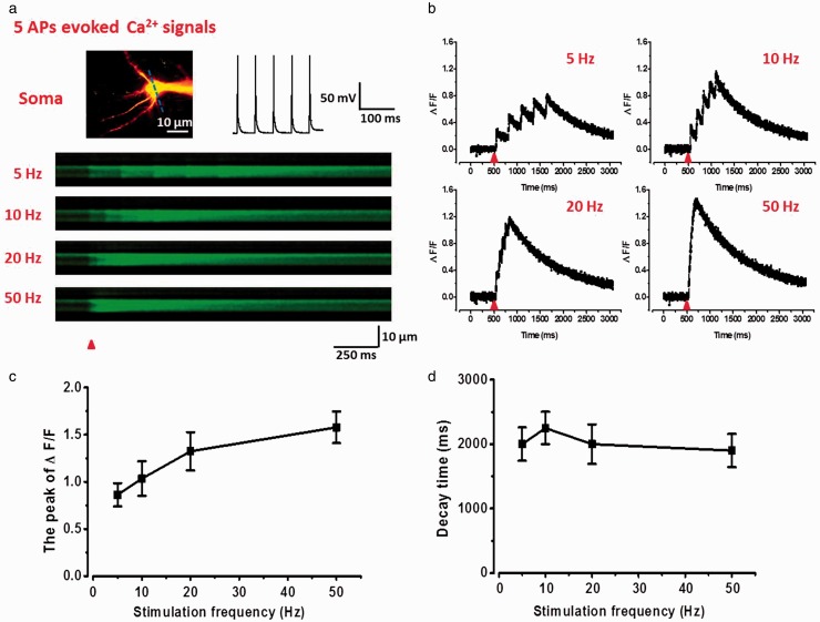
Figure 2.
Summation properties of multi-APs evoked suprathreshold Ca2+ signals in soma of ACC pyramidal neurons. (a) Upper left: two-photon fluorescent image of patched neuron. The blue line indicates the position of the line scan. Upper right: representative current-clamp traces of five spikes action potentials evoked by current injection of 200 pA (5 ms). Below: representative calcium transients images in response to five spikes APs trains at 5, 10, 20, and 50 Hz in soma. (b) Representative waveforms of fluorescence changes (ΔF/F) in response to five spikes APs at 5, 10, 20, and 50 Hz in soma. (c) Summary results showing the peak values of ΔF/F to five spikes APs at different frequencies (n = 6). (d) Summary results showing the decay time of ΔF/F to five spikes APs at different frequencies.
APs: action potentials.

