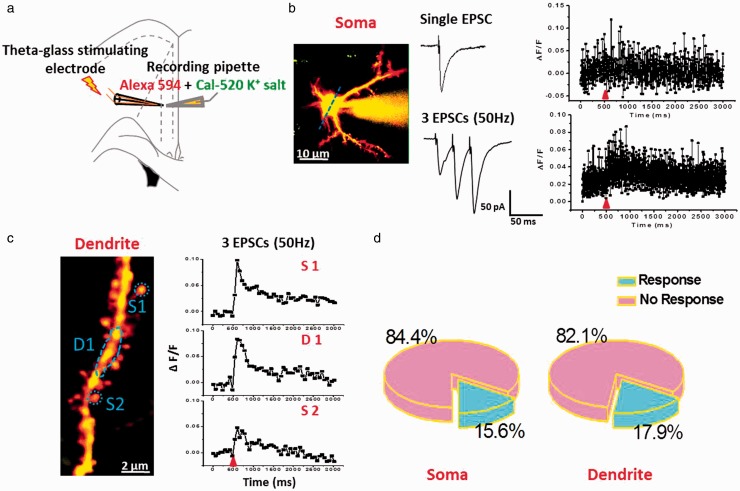
Figure 4.
Electrical stimulation evoked weak subthreshold Ca2+ signals in soma and dendritic spine of ACC pyramidal neurons. (a) Schematic diagram showing the placement of stimulating electrode and recording pipette in the layer II/III pyramidal neurons of the ACC. (b) Left: two-photon fluorescent image of patched active neuron. The blue line indicates the position of the line scan. Middle: representative voltage-clamp traces of single EPSC and three traces EPSCs at 50 Hz induced by local stimulation. Right: associated waveforms of fluorescence change (ΔF/F) in response to single EPSC and three traces EPSCs at 50 Hz in soma. (c) Left: two-photon fluorescent image of dendritic segment containing active spines. Right: waveforms of fluorescence change (ΔF/F) in response to three traces EPSCs at 50 Hz for three active regions (S1: spine1; D1: dendritic region1, and S2: spine2). (d) Pie graph summarizing the active ratio of evoked Ca2+ singles in soma (n = 32) and dendrite containing active spines (n = 28) after three traces EPSCs at 50 Hz stimulation.
EPSCs: excitatory postsynaptic currents.

