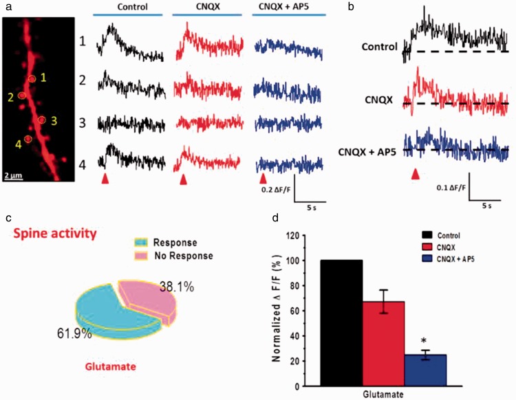
Figure 7.
NMDA receptor-mediated puff-application of Glu evoked Ca2+ signal in spine of ACC pyramidal neurons. (a) Far left, two-photon fluorescent image of a dendritic segment containing active spines. The yellow dashed circles indicate the position of the frame scan. Right, four spines representative calcium transients waveforms of fluorescence changes (ΔF/F) in response to puff-application of 1 mM Glu (10 psi, 100 ms) in the control, CNQX, and CNQX + AP5 conditions, respectively. (b) Average traces of Ca2+ signals (ΔF/F) in responsive spines evoked by puff-application of Glu in the control ACSF, presence of CNQX (20 µM), and AP5 (50 µM), respectively. (c) Pie graph showing the percentage of active spines in the puff-application of Glu. (d) Summary results showing the percentage of Ca2+signals (ΔF/F) in responsive spines in the presence of CNQX and AP5. *P < 0.05, error bars indicated SEM. The amplitudes of Ca2+ signals (ΔF/F) were normalized to control values.

