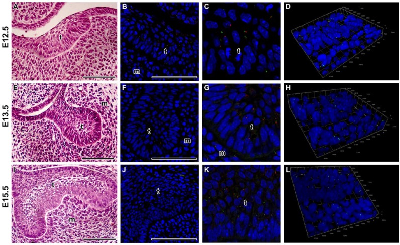
Figure 1.
Localization of primary cilia during early tooth germs in mouse. (A–D) Primary cilia are dispersed through the epithelial thickening. Ciliated cells are also located in the mesenchymal condensation just below the thickening. (E–H) At the bud stage, cilia are situated mostly on the apical surface of the basal epithelial layer. In the central area, there are only a few ciliated cells with shorter cilia. (I–L) Ciliated cells are dispersed through the enamel organ at the early bell stage. (C, G, K) Detail of interface between the dental epithelium and mesenchyme. D, H, L) Three-dimensional view of detail in the area of interface. Primary cilia are labeled by acetylated alpha tubulin (ALEXA488, green) and pericentrin (ALEXA594, red), and nuclei are stained by DRAQ5 (blue). T, tooth germ; m, mesenchyme. Scale bar = 100 µm.

