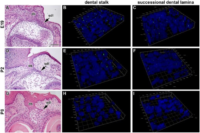
Figure 3.
Three-dimensional analysis of primary cilia directionality in the dental stalk and rudimentary successional dental lamina of mouse. (A, D, G) Lower-power view of the dental stalk and rudimentary successional dental lamina stained by hematoxylin and eosin. (B, E, H) Three-dimensional view of the area of interface between the dental stalk epithelium and mesenchyme. (C, F, I) Three-dimensional view of the area of interface between the successional dental lamina epithelium and mesenchyme. Cilia are short in both areas during late embryonic and postnatal stages and oriented mostly in the rostro-caudal direction in epithelial tissues. The number of cilia decreased with the age of animals. Primary cilia are labeled by acetylated alpha tubulin (ALEXA488, green), and nuclei are stained by DRAQ5 (blue). Ds, dental stalk; sdl, successional dental lamina. Scale bar = 100 µm.

