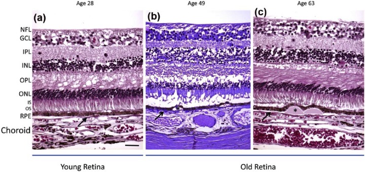
Figure 2.
Histological depiction of young and old retinas. (a) The young retina demonstrates normal retina layers. (b, c) The aged retinas show thinning of the outer nuclear layer (ONL). The aged retinas contain drusen (asterisks) displacing the RPE. The BM is marked with a black arrow. Scale bar: 50 μm. Paraffin sections cut at 4–6 μm.
Source: Reprinted from Ardeljan and Chan.7
NFL: nerve fiber layer; GCL: ganglion cell layer; IPL: inner plexiform layer; INL: inner nuclear layer; OPL: outer plexiform layer; ONL: outer nuclear layer; IS/OS: inner/outer segments of photoreceptors; RPE: retinal pigment epithelium.

