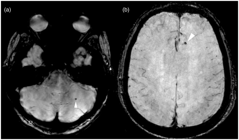
Figure 3.
Mixed distribution of cerebral microbleeds in Alzheimer’s disease patient. (ab) Axial SWI MR images show small, rounded, hypointense cerebral microbleeds in the (a) left cerebellar hemisphere and in the (b) left frontal lobe (arrowhead). SWI: susceptibility-weighted imaging; MR: magnetic resonance.

