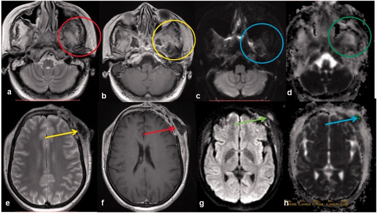
Figure 1.
Skull osteomyelitis: a 35-year-old female with skull bone osteomyelitis. Peripherally enhancing (yellow and red arrows) collections are noted along the left frontal bone extending to involve the skull base, clivus, greater wing and body of sphenoid (red and yellow circles). A breach is noted in the inner and outer skull tables with extension of the collection along the subgaleal region. DW images ((c), (g)) show brightness with low ADC ((d), (h)). DW: diffusion-weighted; ADC: apparent diffusion coefficient.

