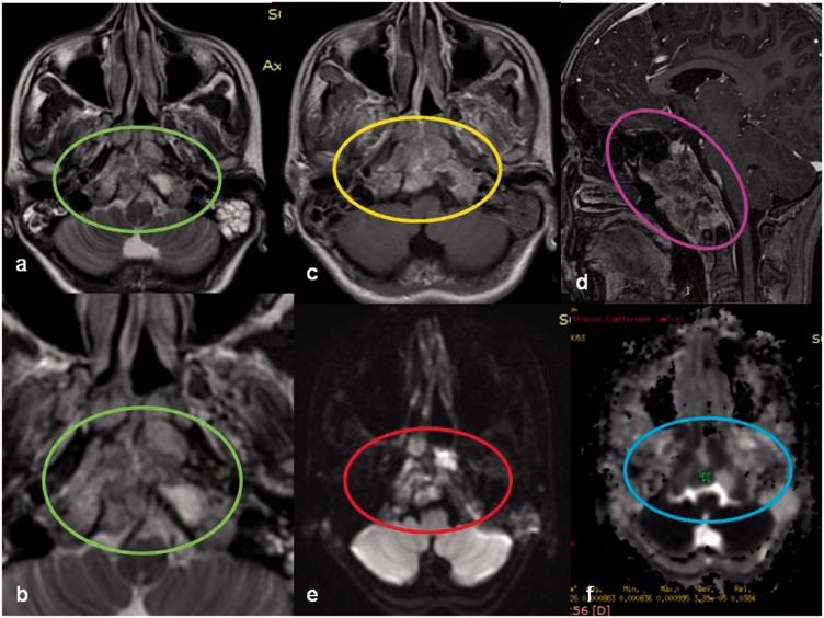
Figure 12.
A clival chordoma in a 12-year-old male child. Large lobulated T2 heterogenously hyperintense ((a), (b)) enhancing ((c), (d)) extraaxial lesion involving the basisphenoid with extension into the sphenoid sinus, nasopharynx, reteropharangeal, and prevertebral space. Lesion shows DWI restriction (red circle) with low ADC (blue circle) suggesting malignant etiology. DWI: diffusion weighted imaging; ADC: apparent diffusion coefficient.

