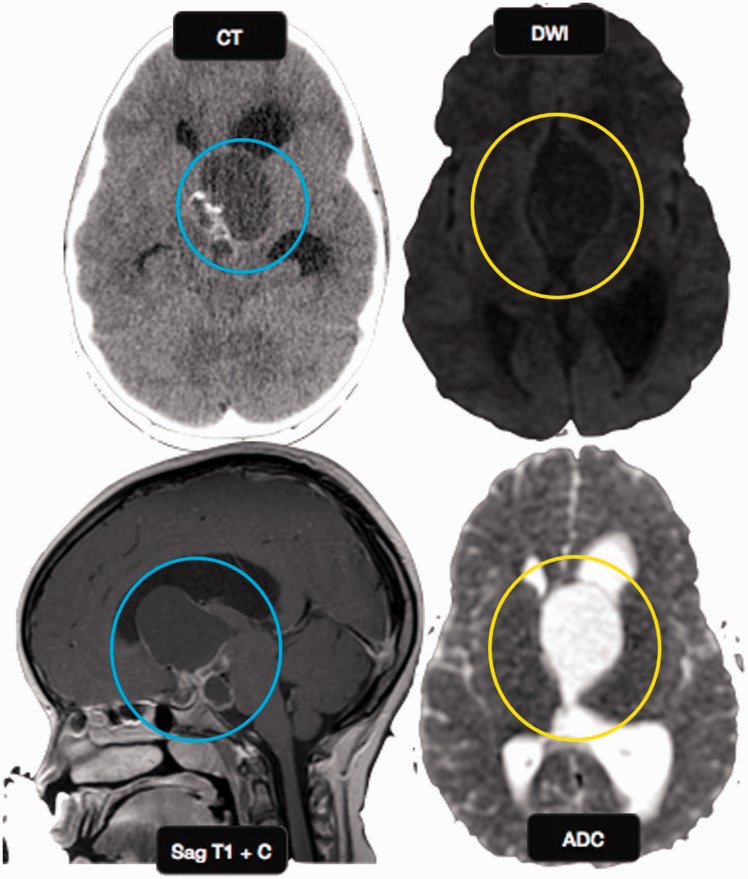
Figure 6.
Craniopharyngioma: an 8-year-old girl with headache, photophobia, and increased urination. Axial CT image shows a cystic suprasellar mass with incomplete rim calcification (blue circle). Post gadolinium T1W sagittal MR image shows rim enhancement. No diffusion restriction was identified on echo planar imaging (yellow circles), suggesting benign etiology. CT: computed tomography; MR: magnetic resonance.

