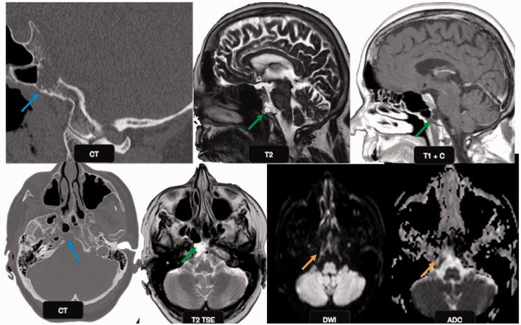
Figure 7.
Ecchordosis physaliphora: a 66-year-old male patient with vertigo and syncope. Magnified sagittal and axial CT scan images show an erosive lesion in the clivus (blue arrows). The lesion appears hypointense on T1 and hyperintense on T2W images (green arrows). No diffusion restriction was identified on echo planar imaging (orange arrows), suggesting benign etiology. This was a case of ecchordosis physaliphora, a benign notochord remnant. CT: computed tomography.

