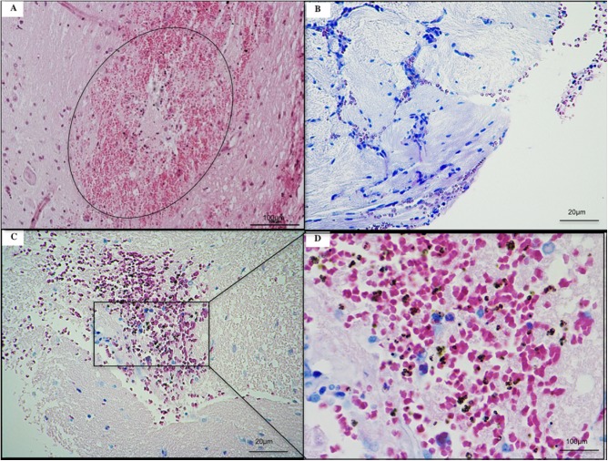
Fig 12. Histological illustrations of cerebral lesions in ECM K173 infected SD rats.
(A) Acute perivascular neuropilar hemorrhage with severe extension (circle). Diffuse intracerebral hemorrhages (B and C) with presence of numerous parasites (D). ECM brains were collected from rats without systemic lavage. Histological slices of ECM brains stained using hematoxylin-eosin.

