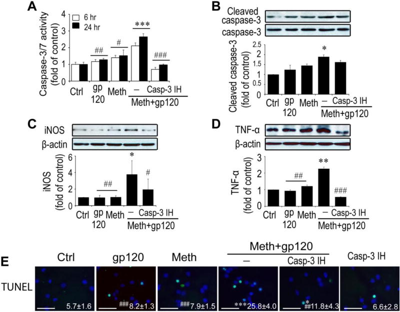
Fig. 4. Meth/gp120-induced neurotoxicity requires caspase-3 signaling.
A. Caspase-3/7 activity was quantified by using caspase-3/7 activity kit (Promega, Madison USA) 6h and 24 after experimental treatments as indicated. Note that Meth and gp120 each alone had minimal effects on caspase-3/7 activity, but they significantly increased caspase-3/7 activity when applied in combination. The Meth+gp120 increase of caspase-3/7 activity was inhibited by Casp-3 IH, demonstrating Meth+gp120 enhancement of microglial caspase-3/7 activity. B, C and D. Western blots illustrate expression of cleaved caspase-3 and its internal control caspase-3 (B), iNOS (C), TNF-α (D), and β-actin of microglia. Increased folds are reflected on bar graphs (lower B, C and D). E. TUNEL staining was visualized by fluorescent microscopy at x100 original magnification. Data represent mean ± SD of values derived from three independent experiments. Statistical significance (*p < 0.05, **p < 0.01, ***p < 0.001 vs ctrl, and # p < 0.05, ##p < 0.01, ###p < 0.001 vs Meth+gp120-treated group. Scale bar equals 50µM.

