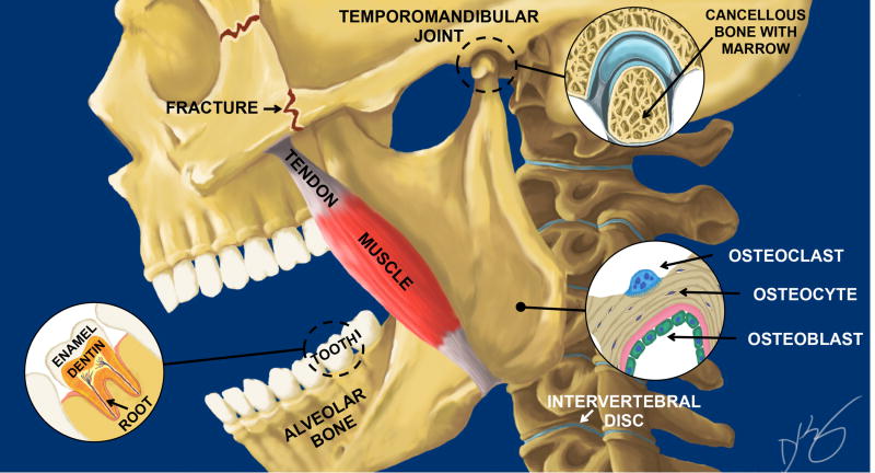
Fig. 1.
Schematic of of a skull depicting key mineralized tissues including a tooth with enamel, dentin and roots, masseter tendon, alveolar bone in the jaw, the temporomandibular joint (TMJ), and the intervertebral disc (IVD). Key cell components of bone are shown in the lower right corner that include the multinuclear osteoclast (blue), the osteocyte (black) and the osteoblast (green). Commonly fractured bones in the face are shown as zigzag lines behind and below the eye socket in the zygomatic arch. Illustration is by David Kirby, co-first author, on the paper in this special edition by Myren et al.

