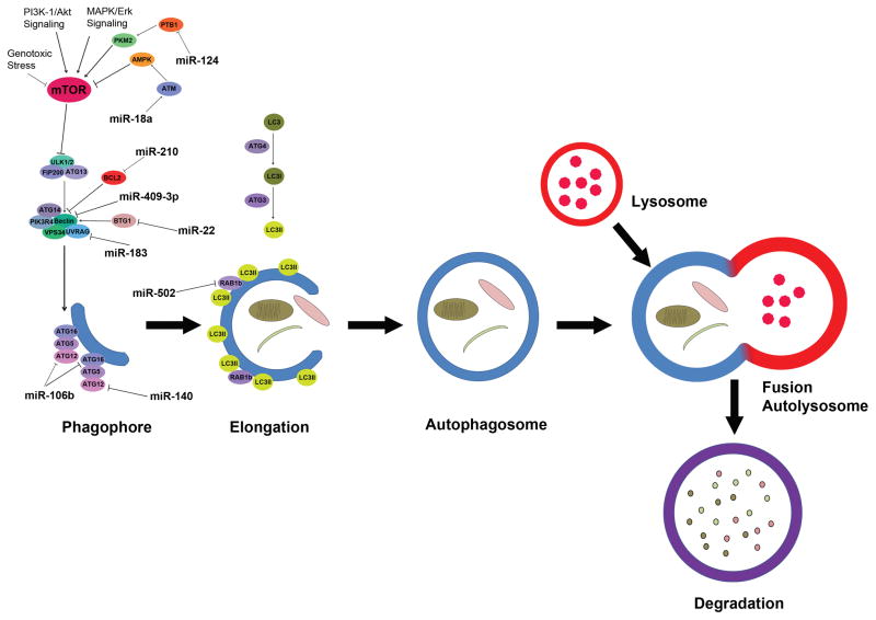
Figure 1.
Schematic illustration of autophagy under stress. The autophagy process is divided into phagophore formation, elongation, autophagosome formation, fusion with lysosome, and degradation. Following the initiation stress signaling through the mTOR kinase, the hypophosphorylated ATG13 is maintained, and the autophagic vesicles are formed from the phagophore/isolation membrane to the autophagosome and autolysosome. ATG-11, -13, -17 and ULK complex mediate the early process followed by Beclin-1-hVPS-34-protein complex. hVPS-34/Beclin-1 complex converts PI to PI3P followed by ATG5-ATG12 conjugation and interaction with ATG16L. Following the LC3 processing events, the nascent complex further undergoes elongation and expansion to form a double membrane autophagosome. The Autolysosome is formed through the fusion of lysosomes docking and fusion with autophagosomes where cargo is degraded to generate amino and fatty acids. miRNAs that involved in colorectal cancer progression and resistance by mediating the initiation and enlongation of autophagy processes are illustrated in the pathway map.

