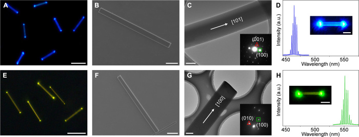
Fig. 2. Preparation of the OPV-A and OPV-B NW lasers.
(A and E) PL images of the OPV-A and OPV-B NWs under UV (330 to 380 nm) excitation. Scale bars, 10 μm. (B and F) SEM images of the typical OPV-A and OPV-B NWs. Scale bars, 5 μm. (C and G) TEM images of the individual OPV-A and OPV-B NWs. Scale bars, 500 nm. Inset: SAED patterns of the NWs. (D and H) Multimode lasing spectra from the OPV-A and OPV-B NWs, excited with a pulsed laser (400 nm). Insets: Corresponding PL images of the OPV-A and OPV-B NWs above the lasing threshold. Scale bars, 5 μm. a.u., arbitrary units.

