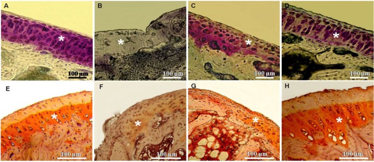
Fig. 3.
Matrix staining of knee joints. A and E) Control group: Strong staining of matrix can be seen. B and F) MIA group: Weak or negative matrix staining is observed. C and G) NCur + MIA group: Moderate matrix staining can be observed. D and H) NCur + MIA group: Strong staining of matrix similar to the control is observed. Asterisks indicate matrix staining. A-D: Toluidine blue staining, E-H: Safranin O staining

