
Fig. 3.
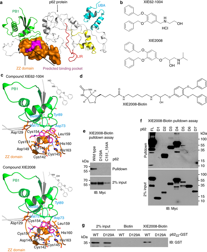
Development of small molecule ligands to p62 ZZ domain using 3D-modeling of p62 and virtual screening. a A 3D-model representing the structure of p62 that shows the predicted binding pocket present in ZZ domain. b The chemical structures of XIE62-1004 and XIE2008. c Docking model of p62 with XIE62-1004 and XIE2008. d The chemical structure of biotinylated XIE2008. e Pulldown assay using biotinylated XIE2008 and myc/His tagged ZZ point mutants expressed in HEK293 cells. 75 μg of total protein was used in pulldown assay, and p62 was detected by immunoblotting analysis using anti-Myc antibody. f Similar to e except that biotinylated XIE2008 pulldown assay was performed using myc/His tagged p62 deletion mutants. g Pulldown assay using biotinylated XIE2008 and 93-residue p62ZZ-GST containing intact ZZ domain